Echocardiogram (ECHO)
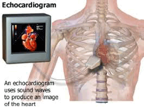
An Echocardiogram uses ultrasound, or harmless sound waves, to obtain valuable information about your heart quickly and efficiently. When our doctors have concerns about the size, shape, or performance of your heart and its valves, they will use an Echocardiogram, or echo.
Echocardiography is a versatile diagnostic tool for detecting heart disease. We provide a comprehensive range of tests, including newer options that can provide 3-D imaging or fit inside the arteries. We use the type that is best suited to your heart and needs.
What Is An Echocardiogram?
Types
The type of Echocardiogram you receive is determined by the potential heart problem that the doctors must investigate. We provide a comprehensive range of echo testing services, including:
- Transthoracic Echocardiogram (TTE): The most common type of Echocardiogram is transthoracic, which is noninvasive and takes place entirely outside your body. A team member applies gel to your chest before scanning your heart with a handheld transducer.
- 3-D echocardiography: While most echocardiography images are flat, our machines can also produce 3-D images. This technology is especially useful for detecting problems with heart valves, replacement heart valves, and the lower left chamber of the heart (left ventricle). We're looking into new ways to use 3-D echo.
- Intracardiac Echocardiogram (ICE): This is a newer type of testing that involves taking images inside your heart. This method is mostly used to monitor treatments that involve inserting thin tubes called catheters into your arteries.
- M-mode Echocardiogram: The most basic type of echocardiography produces an image that looks like a tracing rather than a picture of heart structures. M-mode echo is useful for measuring heart structures such as the heart's pumping chambers, heart size, and heart wall thickness.
- Stress Echocardiogram: An Echocardiogram may be required as part of a comprehensive stress test that intentionally raises your heart rate and blood pressure. We take two sets of images: one at rest and one after a treadmill or stationary bike workout. If your health prevents you from engaging in such physical activity, we will administer a medication that mimics the effects of exercise. This is known as a pharmacologic stress Echocardiogram.
- Transesophageal Echocardiogram (TEE): After sedation, a special ultrasound probe is guided into your mouth and down your esophagus. We can take better images because the esophagus and heart are so close together, and sound waves don't have to travel through skin, muscle, or bone. For some conditions, TEE is a better option. We may also require a higher resolution image of a specific part of the heart. Obesity and lung disease can also interfere with standard echocardiography.
- Doppler Echocardiogram: This technique is used to measure and evaluate the flow of blood through the chambers and valves of the heart. The amount of blood pumped out with each heartbeat is an indication of how well the heart is working. Doppler can also detect abnormal blood flow within the heart, which can indicate a problem with one or more of the four valves or the heart's walls.
What You Can Expect
According to the Best Cardiologist Doctor in Max Hospital Delhi before an Echocardiogram can be performed, some preparation is required. There are also steps to take before and after the procedure. Learn more about what to expect by reading on.
Prior to the Procedure
The doctor will explain the procedure and provide you with the opportunity
to ask questions about it.In most cases, no prior preparation, such as fasting or sedation,
is required.
All medications (prescription and over-the-counter) and herbal supplements should be
reported to your doctor.
Your doctor may request additional preparation based on your medical condition.
Throughout the Procedure
An Echocardiogram can be done as an outpatient procedure or as part of a hospital stay Procedures may differ depending on your condition and your doctor's practices.
This is usually followed by an Echocardiogram:
- You will be asked to take off any jeweler or other objects that could obstruct the procedure. It is possible that you will be required to wear glasses, dentures, or hearing aids.
- You will be asked to remove your clothes and will be given a gown to wear while lying on your left side on a table or bed. Place a pillow or wedge behind your back for extra support.
- You will be hooked up to an ECG monitor, which records your heart's electrical activity and monitors it during the procedure with small, adhesive electrodes. The images displayed on the Echocardiogram monitor will be compared to the ECG tracings that record the electrical activity of the heart.
- The room will be darkened in order for the technologist to see the images on the echo monitor. The technologist will apply warmed gel to the patient's chest before placing the transducer probe on the gel.. As the technologist positions the transducer to obtain the desired image of the heart, you will feel a slight pressure.
- During the test, the technologist will move the transducer probe around and apply varying amounts of pressure to obtain images of various heart locations and structures. The pressure used behind the probe should not be painful.
Following the Procedure
As advised by Best Cardiologist in Saket Delhi unless your doctor advises otherwise, you may resume your normal diet and activities. In general, no special care is needed after an Echocardiogram. Depending on your needs, your doctor may give you additional or alternate instructions after the procedure.